Retinal degeneration is associated with several ocular diseases, like glaucoma or retinal ischemia. As the pathogenic processes of those diseases are not yet completely understood, models are needed to investigate pathologic changes. Furthermore, model systems are necessary to develop new therapeutic approaches and to test novel drugs. Those studies are mostly based on disease-specific animal models, which are bred and euthanized specifically for the experiments. Organ cultures of porcine retina explants, obtained from slaughtered animals for the food industry, offer a good alternative to these animal models. In a prior project, funded by the set Foundation, several degeneration models of the organotypic culture model of the porcine retina were successfully established.
The goal of this project is to investigate, if the cobalt chloride (CoCl2) and the hydrogen peroxide (H2O2) degeneration model can not only be used to analyze the underlying processes of retinal degeneration (Kuehn et al. 2017; Hurst et al. 2017), but also to screen for therapeutics and to test therapeutic treatments, such as hypothermia. Therefore, a possibly protective effect of different therapeutic approaches, like hypothermia or inhibitors, will be investigated in detail. This is of eminent importance since the successful testing of such therapies is crucial to get the model approved as an animal replacement method.
Once again porcine eyes from the local abattoir are used for these studies. Therefore, no animals are killed exclusively for these experiments, but waste products from the food industry are used. This study aims at completely replacing animal experiments in the first test period of therapy screenings (Replace). Additionally, the size of the porcine eyes offers the crucial advantage to prepare four comparable samples from one eye and thereby the possibility to correlate the results of different methods statistically more accurate. In comparison to animal models, the size of a rat or mouse eye is only sufficient for one sample each. The number of animals can be considerably decreased as compared to rat or mouse eyes (Reduce).
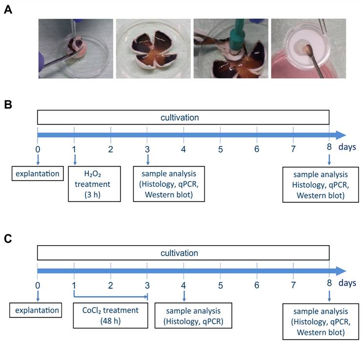
Fig. 1: Porcine retinal organ culture and test procedures for the degeneration models:
A) For the preparation of the porcine retinal organ culture the front part of the eye is cut away, a cloverleaf structure is formed and the retina is punched out and placed on a filter. The retina can then be cultivated in Neurobasal-A medium. B) The timeline for the H2O2 degeneration model is shown. The retinal explants were treated with H2O2 for three hours on day 1. After three and eight days the samples were analyzed histologically and using qRT-PCR and Western blot analysis. C) The procedure for the CoCl2 degeneration model is also displayed. There, the retinal explants were treated with CoCl2 for 48 hours from day one up to day three. After four and eight days the samples were analyzed histologically and using qRT-PCR and Western blot analysis.
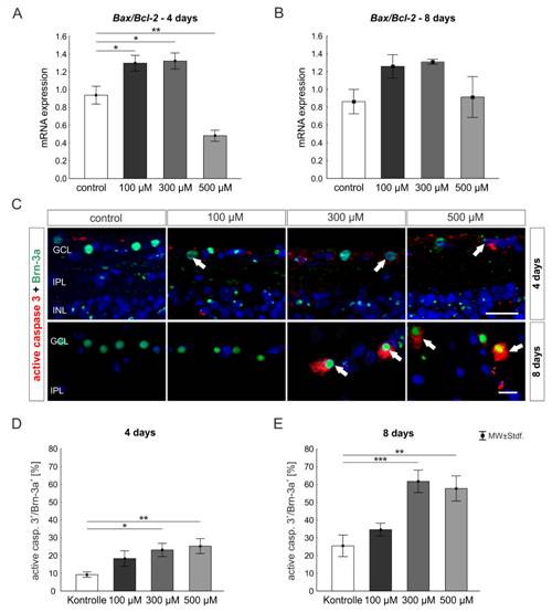
Fig. 1: Activation of different cell death mechanisms in porcine retinal organ cultures treated with cobalt chloride:
A) The Bax/Bcl-2 ratio was analyzed on mRNA level after four days. Bax/Bcl-2 mRNA was increased only in the samples treated with 100 and 300 µM CoCl2 (both: p<0.05). The treatment with 500 µM even led to a downregulation of this ratio (p<0.01). B) After eight days, no difference in the Bax/Bcl-2 mRNA level could be detected between the groups. C) Immunohistological staining of the ganglion cell marker Brn-3a (green) in combination with activated caspase 3 (red) and a cell nucleus marker (DAPI, blue) were shown. D) After four days, more apoptotic ganglion cells were noted in the samples treated with 300 (p<0.05) and 500 µM CoCl2 (p<0.01). E) In both groups the amount of apoptotic ganglion cells further increased after eight days (300µM: p<0.001; 500µM: p<0.01). Scale = 20µm. Abbreviations: GCL = ganglion cell layer; IPL = inner plexiform layer (Kuehn et al. 2017).
Institutions
Research lab, University Eye Clinic, Knappschaftskrankenhaus, Ruhr-University Bochum
In der Schornau 23-25, 44892 Bochum
Research lab, University Eye Clinic Tübingen
Elfriede-Aulhorn-Str. 7, 72076 Tübingen
Duration
01/2017 - 06/2019

 English