Current drug development is usually based on the principle to test new substances in two-dimensional (2D) cell culture and subsequently characterize them in an animal model. This procedure, however, has several drawbacks. Cells in a 2D cell culture do not represent the physiology of cells in a three-dimensional (3D) tissue, as they, for example, differ in their gene expression patterns. Animal models have the disadvantage that the animal cells differ from human cells. In addition, in vivo experiments are usually associated with unethical suffering of the animals.
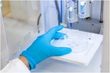
To solve these problems, 3D organ models are being developed. In these models, cells are cultivated in the natural 3D environment. It is possible to employ human cells, and the use of animal models can be avoided. Great advancements were made with the development of additive manufacturing (3D bioprinting) technologies.
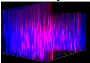
The aim of this project was to create humanized organ models using sophisticated bioprinting technologies. First a lung model was printed. The composition of the bioink was optimized. Finally, an alginate/gelatin/Matrigel mixture was used to create a model in which the cells had a good three-dimensional distribution and high viability. Finally, the lung model was infected with influenza A viruses. The viruses replicated and triggered an immune response.
A critical point of the lung model from an animal welfare perspective is the use of Matrigel, an extracellular matrix derived from sarcomas in mice. In a further step, we replaced Matrigel with an extracellular matrix from a human donor. This material was used to print a liver model that was infected with human adenoviruses. These viruses constitute a great danger for immunocompromised patients after organ transplantation. As with the influenza viruses in the lung model, the adenoviruses replicated efficiently in the printed liver model.
In summary, a liver and a lung model could be generated by 3D bioprinting. According to our research, printed organ models were infected with viruses for the first time in the published literature. The humanized models can now be used to develop new antiviral strategies without animal experiments.
Publikations and Awards:
The project has resulted in two primary papers and a review article. The work was honored with the Prize of the State of Berlin for the development of alternatives to animal experiments in research and teaching awarded to Dr. Johanna Berg and Prof. Dr. Jens Kurreck.
Hiller, T.; Berg, J.; Elomaa, L.; Rohrs, V.; Ullah, I.; Schaar, K.; Dietrich, A.C.; Al-Zeer, M.A.; Kurtz, A.; Hocke, A.C.; Hippenstiel, S.; Fechner, H.; Weinhart, M.; Kurreck, J. (2018) Generation of a 3D Liver Model Comprising Human Extracellular Matrix in an Alginate/Gelatin-Based Bioink by Extrusion Bioprinting for Infection and Transduction Studies. Int. J. Mol. Sci., 19.
Berg, J.; Hiller, T.; Kissner, M.S.; Qazi, T.H.; Duda, G.N.; Hocke, A.C.; Hippenstiel, S.; Elomaa, L.; Weinhart, M.; Fahrenson, C.; Kurreck, J. (2018) Optimization of cell-laden bioinks for 3D bioprinting and efficient infection with influenza A virus. Sci. Rep., 8, 13877.
Weinhart, M., Hocke, A., Hippenstiel, S., Kurreck, J. and Hedtrich, S. (2019) 3D organ models-Revolution in pharmacological research? Pharmacol. Res., 139, 446-451.
Institution
Prof. Dr. Jens Kurreck
Technische Universität Berlin
Institut für Biotechnologie, Fachgebiet Angewandte Biochemie, TIB 4/3-2
Gustav-Meyer-Allee 25
13355 Berlin, Germany
Prof. Dr. Hartmut Schwandt
Technische Universität Berlin
Institut für Mathematik, 3D Labor, MA 6-4
Straße des 17. Juni 136
10623 Berlin, Germany


 English


