
Ex vivo liver model produced using 3D printing
Organ models produced using 3D printing can help to develop new pharmacological substances and assess their efficacy and toxicity without the need for animal testing.
Current drug development is generally based on the principle of first testing new substances in two-dimensional (2D) cell cultures and then characterising them in more detail using animal models. However, this approach has several shortcomings. Cells in a 2D cell culture do not generally represent the physiology of cells in a three-dimensional (3D) cell assembly; among other things, gene expression differs. Animal models have the disadvantage, on the one hand, that animal cells differ from human cells; on the other hand, in vivo models are generally associated with unacceptable suffering of the experimental animals. The aim of this project was to create humanised organ models using bioprinting. First, a lung model was printed. In doing so, the composition of the bio-ink was optimised. Ultimately, a model was produced using a mixture of alginate, gelatine and Matrigel, in which the cells exhibited good three-dimensional distribution and high viability. Finally, the lung model was infected with influenza A viruses. The viruses replicated and triggered an immune response.
From an animal welfare perspective, a key concern with the lung model is the use of Matrigel, an extracellular matrix derived from mouse sarcomas. We therefore replaced Matrigel with an extracellular matrix from a human donor. This enabled us to 3D-print a liver model that was infected with human adenoviruses. These viruses pose a major risk to immunocompromised patients, for example following an organ transplant. As with the influenza viruses in the lung model, the adenoviruses replicated efficiently in the printed liver model. Overall, the project succeeded in generating a liver and a lung model via 3D bioprinting. According to our review of the published literature, this marks the first time that printed organ models have been infected with viruses. The humanised models can now be used to develop new antiviral strategies without the need for animal testing.
The project has resulted in two original papers and a review article. The work was recognised with the State of Berlin’s award for the promotion of alternative and complementary methods to animal testing in research and teaching, presented to Dr Johanna Berg and Prof. Dr Jens Kurreck.
Publikationen und Preise:
Aus dem Projekt sind zwei Primärarbeiten und ein Übersichtsartikel hervorgegangen. Die Arbeiten wurden mit dem Preis des Landes Berlin zur Förderung von Ersatz- und Ergänzungsmethoden für Tierversuche in Forschung und Lehre an Dr. Johanna Berg und Prof. Dr. Jens Kurreck gewürdigt.
Hiller, T.; Berg, J.; Elomaa, L.; Rohrs, V.; Ullah, I.; Schaar, K.; Dietrich, A.C.; Al-Zeer, M.A.; Kurtz, A.; Hocke, A.C.; Hippenstiel, S.; Fechner, H.; Weinhart, M.; Kurreck, J. (2018) Generation of a 3D Liver Model Comprising Human Extracellular Matrix in an Alginate/Gelatin-Based Bioink by Extrusion Bioprinting for Infection and Transduction Studies. Int. J. Mol. Sci., 19.
Berg, J.; Hiller, T.; Kissner, M.S.; Qazi, T.H.; Duda, G.N.; Hocke, A.C.; Hippenstiel, S.; Elomaa, L.; Weinhart, M.; Fahrenson, C.; Kurreck, J. (2018) Optimization of cell-laden bioinks for 3D bioprinting and efficient infection with influenza A virus. Sci. Rep., 8, 13877.
Weinhart, M., Hocke, A., Hippenstiel, S., Kurreck, J. and Hedtrich, S. (2019) 3D organ models-Revolution in pharmacological research? Pharmacol. Res., 139, 446-451.
Project management
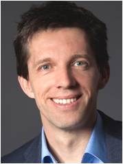
Prof. Dr. Jens Kurreck
Studium der Biochemie und Philosophie an der Freien Universität (FU) Berlin. Dissertation zur Primärreaktion der Photosynthese an der Technischen Universität (TU) Berlin, gefolgt von einem Postdoktoranden-Aufenthalt an der Arizona State University, Tempe, USA. Habilitation zu medizinischen Anwendungen von RNA-Technologien an der FU Berlin. 2007-2009 Professor an der Universität Stuttgart, seit 2009 W3-Professor an der TU Berlin. Arbeitsschwerpunkte: RNA-Technologien, Virale Vektoren, antivirale Strategien, 3D-Organmodelle.
Prof. Dr. Hartmut Schwandt
Studium der Mathematik, Informatik und Betriebswirtschaftslehre an der Technischen Universität Berlin (TUB). Promotion und Habilitation in Numerischer Mathematik/Computermathematik an der TUB. Seit 1982 IT-Leiter am Institut für Mathematik, seit 2007 auch der IT der Fakultät für Mathematik und Naturwissenschaften, seit 1998 apl. Professor für Numerische Mathematik, 2005 Gründung des 3D-Labors an der TUB. Arbeitsschwerpunkte: 3D-Technologien und Anwendungen, mathematische und 3D-Verfahren für Anwendungen in der Verkehrsforschung, Computermathematik, Parallele Numerik.
Cooperation
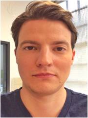
M.Sc. Thomas Hiller
Biotechnologiestudium an der Beuth Hochschule für Technik in Berlin. Abschluss 2012 mit einem M.Sc. Im Anschluss (2012-2014) wissenschaftlicher Angestellter in der experimentellen Chirurgie sowie dem Berlin-Brandenburger Centrum für regenerative Therapien an der Charité Berlin. Arbeitsschwerpunkt: Entwicklung und Etablierung eines oxidativen Stressmodells in primären Hepatozyten des Menschen und der Ratte in einem 3D-Leberbioreaktor. Seit 2014 Doktorand in der Arbeitsgruppe von Jens Kurreck.
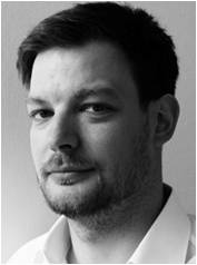
Dipl.-Ing. Ben Jastram
Architekturstudium an der Universität der Künste Berlin. Abschluss 2009 als Diplom-Ingenieur. Im Anschluss (2009-2010) wissenschaftlicher Mitarbeiter im Projekt „Digital Processing for Academics“ am 3D-Labor der TUB für die Vermittlung von 3D-Messtechnik und 3D-Fertigungstechnik. Seit 2010 Wissenschaftlicher Angestellter. In den Projekten „Rapid Manufacturing in der Medizintechnik und anderen Bereichen“ verantwortlich für die Entwicklung von Maschinen- und Verfahrenstechnik für die Herstellung von 3D-Scaffolds und bei „Herstellung vitaler Herzklappen unter Verwendung von Tissue Engineering und 3D-Druck (3D-TERM)“ verantwortlich für die Prozessentwicklung zur Herstellung von biopolymerbasierten Scaffolds.