
Bioprinting of a 3D tumor model
A tumor is to be printed in a human tumor microenvironment in order to investigate the efficacy, specificity and toxicity of new cancer drugs.
The transferability of results from animal experiments to humans is particularly poor in the field of cancer research. Around 97% of all oncology drug candidates that have proven to be efficient and safe in preclinical studies in animal models fail in human clinical trials. One important reason for the low chance of success is the species-specific differences between animals and humans. At the same time, in vivo trials in cancer research are particularly stressful for the animals.
In the funded project, therefore, human tumor models are to be generated by means of bioprinting. In this process, a tumor is embedded in a tumor microenvironment consisting of healthy human cells (Fig. 1).

This allows to study the very important interactions between the tumor and its surrounding cells. As an example, a tumor consisting of neuroblastoma cells surrounded by kidney cells will be printed. The kidney is an occasional, albeit rare, site for metastases from neuroblastoma.
In a further step, the tumor model is treated with various substances. In preliminary experiments, we have already shown that the anticancer drug panobinostat selectively kills the cancer cells by inducing apoptosis, while the healthy cells of the tumor environment remain unaffected (Fig. 2).

In order to reproduce the natural physiology of tissues, it is important to vascularize the in vitro organ models and perfuse them with a media flow. Here, bioprinting offers the possibility of integrating channel structures, which represent natural blood vessels, into the 3D constructs. As part of the project, a channel was successfully implemented in a model of a lung tumor. The model was connected to a media stream (Fig. 3). This increased the viability of the cells, and a tested cancer drug worked better under these dynamic conditions than under static cultivation.

A further developed version of the tumor model consists of a cancerous core located in an environment of healthy cells. This model can be used to study both, the efficiency of new cancer drugs and their side effects on surrounding tissue.
Publikations:
Wu, D.; Berg, J.; Arlt, B.; Röhrs, V.; Al-Zeer, M.A.; Deubzer, H.E.; Kurreck, J. Bioprinted Cancer Model of Neuroblastoma in a Renal Microenvironment as an Efficiently Applicable Drug Testing Platform. Int. J. Mol. Sci. 2022, 23, 122. https:// doi.org/10.3390/ijms23010122
Mei, Y.; Wu, D.; Berg, J.; Tolksdorf, B.; Roehrs, V.; Kurreck, A.; Hiller, T.; Kurreck, J., Generation of a Perfusable 3D Lung Cancer Model by Digital Light Processing. Int. J. Mol. Sci. 2023, 24, 6071.
Wu, D.; Pang, S.; Berg, J.; Mei, Y., Ali, A.S.M.; Röhrs, V.; Tolksdorf, B.; Hagenbuchner, J.; Ausserlechner, M. J.; Deubzer, H.E.; Gurlo, A.; Kurreck, J. Bioprinting of perfusable vascularized organ models for drug development via sacrificial-free direct ink writing. (Submitted)
Project management
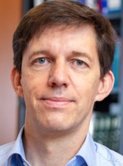
Prof. Dr. Jens Kurreck
Study of biochemistry at Freie Universität Berlin, doctorate at TU Berlin. Postdoctoral fellow at Arizona State University in Tempe, USA. From 2007-2009 professor at the University of Stuttgart. Professor of applied biochemistry at TU Berlin since 2009. Co-spokesperson of the Einstein Center 3R for Animal Experimentation Alternatives. Main research interests: 3D organ models, RNA technologies and antiviral strategies.
Cooperation
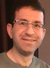
Dr. Munir Al-Zeer
Graduation from Humboldt-Universität zu Berlin and Max Planck Institute for Infection Biology in 2009. PhD under the supervision of Prof. Thomas Meyer. Since 2017 research associate at the Department of Applied Biochemistry, Technische Universität (TU) Berlin. Works on questions of particular clinical relevance, the relationship between chronic bacterial infections, inflammation, and cancer using 3D printing and organoid technologies as an alternative to animal testing.
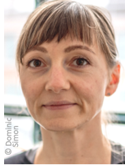
Dr. Johanna Berg
Studied biology at Freie Universität Berlin and Christian-Albrecht-Universität zu Kiel. PhD thesis on the use of human lung tissue in infection research at Charité - Universitätsmedizin Berlin in 2016. Since then postdoctoral researcher at the Department of Applied Biochemistry at TU Berlin. She is working on the bioprinting of organ models for infection studies. In 2019, she, together with Jens Kurreck, received the Award of the State of Berlin for the Development of Replacement and Complementary Methods for Animal Experiments.