
Analysis of toxicity induced cell degeneration in an organotypic porcine retinal culture
For studies of retinal degeneration eyes from pigs slaughtered for food industry could be used instead of the small rodents used so far.
The phenomenon of retinal degeneration occurs in many eye diseases, like glaucoma, Retinitis pigmentosa or retinal ischemia. Since the formation of these diseases is not fully understood yet, reliable models are needed to determine the pathological alterations. To date, these studies were based on acute disease induced animal models, which are bred and killed just for these experiments. Organotypic cultures of retinal explants, obtained from eyes from pigs slaughtered for the food industry, offer a good alternative. Retinal organ cultures are in-vitro-models with a high significance, since the cells are still in their natural cell-organization. In addition, the pig eye is more similar to the human eye in size and morphology than the most common laboratory animals, like rats, mice and rabbits. The goal of this project is to modify this model through the use of in-vivo stressors so it can be used as a screening-model for novel therapy options.
During this project the effect of three stressors on the loss of neuronal cells in the retina, especially ganglion cells, will be evaluated. Therefore, retinal explants, reproducible in size and shape, will be analyzed histologically in regard to possible alterations of ganglion cells but also the other structures of the retina are evaluated.
Apoptotic and immunologic processes play an important role during degeneration. To quantify the alterations of the different processes, like caspase-pathway or glia cells, they will be analyzed using quantitative real-time PCR on mRNA level and using Western-blot on protein level. The localization of the cell alterations in the retina is possible with histological evaluation. When the damage patterns and alterations induced by the stressors are known the optimal stressor-dose for the therapy-studies can be determined. During the therapy-studies two possible neuroprotectants will be tested against these stressors. The same methods as described before will be used for these studies. The RNA-expression (quantitative real-time PCR) and the protein-expression (histology and Western-blot) will be determined, to find out how they are altered by the stressors. This way, the effect of the neuroprotectants on the degenerating retinas can be evaluated.
This model can be seen as a model-system for pathological alterations during retinal diseases, e.g. glaucoma. It can be used for future studies testing therapeutic substances and for studies analyzing pathomechnisms or mode of actions and therefore contribute to lower numbers of research animals. This model can therefore replace animal experiments (Replace). Based on the size of the pig eyes they have the great advantage that four comparable samples can be taken from one retina. Using mice or rats the size of the eye and therefore the volume of the material (protein, DNA and RNA) would only be sufficient for one sample. The number of animals can, compared to mice and rats, be reduced by factor four (Reduce). Through the use of a porcine organ-culture-model for screening of therapeutic substances the doses of these substances can be optimized, before they are used in an animal model in-vivo (Refine).
Project management
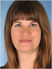
PD Dr. Stephanie Joachim
Studies in human medicine in Ulm and Mainz 1996-2004, Thesis 2006. Post-Doc Fellowship Alcon Laboratories Inc., Fort Worth, TX, USA 2004-2005. Post-Doc at the Experimental Ophthalmology at the University Eye Clinic, Universitymedicine Mainz 2006-2010. Since 2010 Head oft he research lab at the University Eye Clinic at the Ruhr-University Bochum at the Knappschaftskrankenhaus. Habilitation in Experimental Ophthalmology 2014.
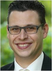
Dr. Sven Schnichels
Studies of biology in Hohenheim 2000-2005, Diploma in biology 2005. PhD studies at the University Tübingen 2006-2010. Post-Doc at the University Eye Hospital Tübingen 2009-2011. Laboratory head for preclinical research 2011-2013. Laboratory head at the University Eye Hospital Tübingen since 2013.
Cooperation
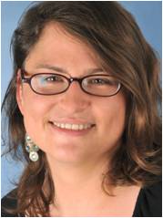
Sandra Kühn, Dipl. Biochem.
Studies in biochemistry at the Ernst-Moritz-Arndt University Greifswald 2005-2010, Diploma in Biochemistry 2010. Since 2011 PhD student at the research lab, University Eye Clinic, Knappschaftskrankenhaus, Ruhr-University Bochum.
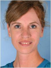
Gesa Stute, B.Sc.
Studies in Biology at the Ruhr-University Bochum 2007-2012, Bachelor of Science 2012. Since 2013 technical assistant at the research lab, University Eye Clinic, Knappschaftskrankenhaus, Ruhr-University Bochum.